Products
Veterinary Equipment
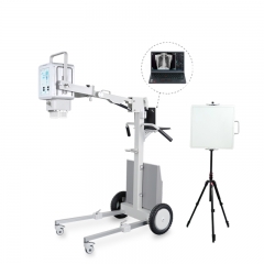
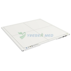
Veterinary X-ray Machine
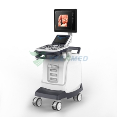
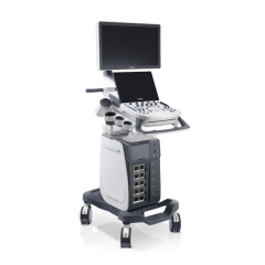
Veterinary Ultrasound Scanner
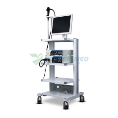
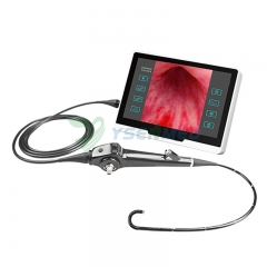
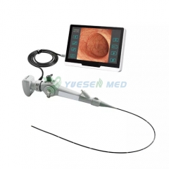
Veterinary Endoscope System
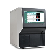
Veterinary Lab Equipment
Veterinary Cages
Veterinary Operating Table
Veterinary Anesthesia & Ventilator
Veterinary Patient Monitor
Veterinary Grooming Equipment
Veterinary Treadmill
Veterinary Surgery Warming System
Veterinary ECG
Veterinary Weighing Scale
Veterinary Infusion Table
Veterinary Infusion & Syring Pump
Veterinary Anatomy Table
Veterinary Surgical Instrument
Veterinary Treatment Table
Veterinary ICU Incubator
Veterinary Electrosurgical Generator
Veterinary lasers
Veterinary Dental Equipment
Veterinary Instrument Trolley
Veterinary Stretcher
Veterinary Drill and Saw
Veterinary Restraint Unit
Veterinary ENT Equipment
Veterinary PCR Systems
Veterinary Hemodialysis
Operating Room Equipment
Anesthesia Machine
Medical Ventilator
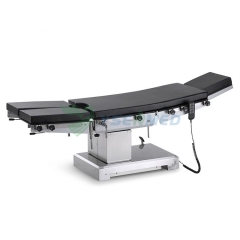
Operating Table
Operation Lamp
Electrosurgical Unit
Patient Monitor
Defibrillator
Surgical Instrument
Ultrasonic Scalpel
ECG Machine
Oxygen Machine
Oximeter
Infusion Pump
Syringe Pump
Surgical Pendant
Electric Dermatome
Electric Drill & Saw
Suction Unit
Spirometer
Gastric Lavage Machine
Automatic Tourniquet
Ozone Therepy Machine
Vein Finder
EEG
Operating Microscope
Laboratory Equipment
Hematology Analyzer
Biochemistry Analyzer
Blood Gas Analyzer / POCT
Immunoassay Analyzer
Urine Analyzer
Electrolyte Analyzer
Elisa Reader & Washer
Coagulation Analyzer
Protein Analyzer
Centrifuge
Microscope
Incubator
PCR
Feces Analyzer
Electrophoresis
Plasma Equipment
Hemoglobin Analyzer
ESR Analyzer
Multi-Gas Analyzer
Biosafety Cabinet
Flow Cytometry
UV Transilluminator
Gel Imaging System
Carbon Dioxide Analyzer
Dry Bath
Uric Acid Analyzer
Blood lipid Analyzer
Renal Function Analyzer
UV Spectrophotometer
Plasma Thawing Machine
Laboratory Pure Water Machine
Moisture Analyzer
Water quality analyzer
Heating Mantle
Digital Rotary Evaporator
Helicobacter Pylori Detector
Chromatography
ENT Instrument
ENT Treatment Unit
OCT
ENT Diagnostic Device
Anesthesia Laryngoscope
Slit Lamp
Surgical Microscopes
Ophthalmic Scanners
Surgical Headlights
Fudus Camera
Refractometer
Tenonometer
Auto Perimeter
Eyesight Screening Tester
Phoroptor
Corneal Topographer
Lens Meter
Frame Heater
Lens Photochromic Tester
Lens Punching Machine
Lens Thickness Meter
Lens Groover
Lens Polisher
Screw Extractor
Simulated Eyes
Three-hole Driller
Trial Frame
LED Vision Chart
Vision Chart Projector
Pupil Distance Meter
Glasses Mounting Tools
Glasses Ultrasonic Cleaner
Trial Lens Set
LCD Chart Monitor
Stress Meter
Layout Blocker
Lens Edger
Audiometer
Dry Eye Analyzer
Hearing Aid